Dr. Vidhi Majithia - Best Private Hospital in Dubai Al Mankhool | IMH Dubai

Cataract

What is a Cataract?

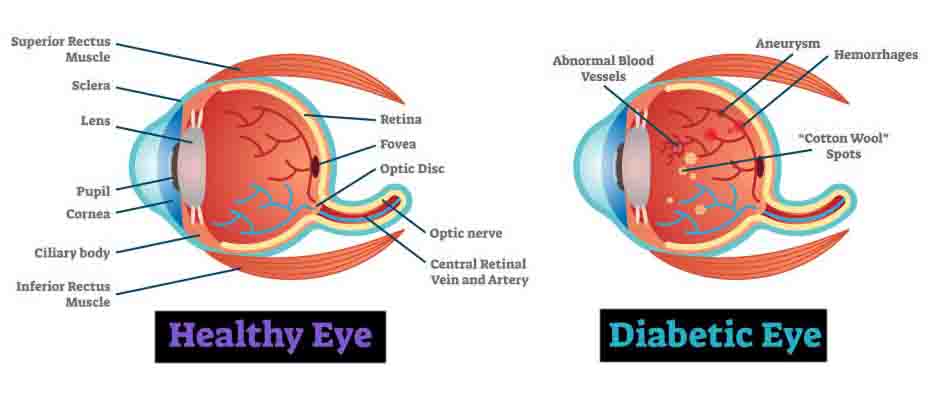
A cataract is a clouding of the eye’s lens, which blocks or changes the passage of light into the eye. The lens of the eye is located behind the pupil and the colored iris, and is normally transparent. The lens helps to focus images onto the retina – which transmits the images to the brain.

Am I at risk for developing cataracts?

The exact cause of a cataract is unknown. Most often, a cataract is part of getting older. As you age, you are at greater risk of developing a cataract. There are also several possible risk factors for cataracts, such as:
• Intense heat or long-term exposure to UV rays from the sun
• Certain diseases, such as diabetes
• Inflammation in the eye
• Hereditary influences
• Events before birth, such as German measles in the mother
• Long-term steroid use
• Eye injuries
• Eye diseases
• Smoking

What are the symptoms of a cataract?

Generally, a cataract does not cause pain, redness or tears. The following problems may indicate that you have a cataract:
• You have blurred vision, double vision, ghost images, or the sense of a “film” over your eyes.
• Lights seem too dim for reading or close-up work, or you are “dazzled” by strong light.
• You change eyeglass prescriptions often and the change does not seem to help your vision.
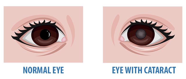
• You may also be able to see the cataract in your eye. It may look like a milky or yellowish spot in your pupil.

Why do cataracts form?

Congenital cataract

Cataracts are probably caused by changes related to aging. Throughout our lives, our bodies replace old cells with new ones. As we grow older, the old cells in our eye’s lens build up and block light as it tries to pass through. The end result is cloudy vision.

As mentioned earlier, besides getting older, other factors may cause cataracts to form. Each of these factors lead to a metabolic pathway which leads to cloudiness of the otherwise transparent lens..

How will my eye doctor (Ophthalmologist) check for cataracts?

Everyone who gets a cataract experiences it differently. But a person with a cataract commonly experiences cloudy or blurry vision. Lights may cause a glare, seem too dim or seem too bright. It may be hard to read or drive, especially at night. If you have a cataract, you may see halos around lights, such as car headlights, that make it hard to focus clearly. Colors may not seem as bright as they used to be. Or you may have to change your eyeglass prescription often.

If you notice any of these changes, make an appointment to see your eye doctor.
If you have a cataract, you may have symptoms that are similar to those of other eye diseases. Only your eye doctor can tell you for sure what’s wrong.

To find out if you have cataracts, your eye doctor will want to:
• Find out your general medical history
• Find out your specific eye history, including problems and symptoms
• Test your vision (visual acuity)
• Test your side vision (peripheral vision)
• Test your eye movement
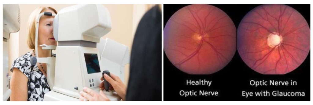
• Test you for glaucoma (by measuring the eye’s internal pressure)
• Do a microscopic exam of the front of the eye (using something called a slit lamp) to assess the density of the cataract and how it interferes with light passing through the lens
• Widen (dilate) the pupils of your eyes to examine the retina, the optic nerve (which carries visual messages from the retina to the brain) and the macula (responsible for the best part of central vision)
• Test you to see how glare affects your vision
After this exam, your eye doctor will determine whether you have cataracts, how much they interfere with your vision, whether surgery would help, and what types of treatment and lens replacements are best for you.

Glaucoma – the ‘sneak thief of sight’

Glaucoma is a serious eye condition that can cause blindness. It damages the optic nerve, which carries information from your eyes to the visual center in your brain. This damage can result in permanent vision loss. The most common type of glaucoma has no early warning signs and can only be detected during a comprehensive eye exam. If Undetected and untreated glaucoma first causes peripheral vision loss and eventually can lead to blindness. Glaucoma is caused by higher than normal pressure inside the eye. By the time you notice vision loss from glaucoma, it’s too late. The lost vision cannot be restored, and it’s very likely you may experience additional vision loss, even after glaucoma treatment begins.

What are the Types of Glaucoma?

  1. Angle Closure Glaucoma : The drainage angle is too narrow or begins to close, impeding the normal outflow of aqueous humor.
  2. Congenital Glaucoma : Some children are born with a defect in the drainage angle that prevents the aqueous fluid from exiting the eye normally.
  3. Secondary glaucoma : Develops due to complications of certain conditions (eg: Trauma, Diabetes), Side effects of medications or because of other eye conditions.

What are the Symptoms of Glaucoma?

• Blurred vision
• Seeing colored rings around lights
• Eye pain
• Headache
• Light sensitivity
• Redness in Your Eye
• Nausea and vomiting in later stages

How is Glaucoma Diagnosed?

• Detailed medical History (including family history)
• Tonometry –Eye pressure check
• Pachymetry – Corneal thickness check
• Perimetry – Visual field test
• Optical Coherence Tomography- Optic Nerve scan

Who is at the risk of Glaucoma?

• Age- People over 60 are at increased risk of glaucoma
• Eye Problems-Chronic eye inflammation, Trauma, thin cornea
• Family History
• Medical History-High Blood pressure, Heart disease, Diabetes
• Use of Certain Medicine-Corticosteroids for extended periods

How is glaucoma Treated?

• Medications- mild to moderate stage
• Surgery- severe stage

Can Glaucoma be prevented?

Glaucoma can’t be prevented. But it is still important to catch it early so treatment can begin in the early phase that will help prevent it from worsening

Diabetic Retinopathy

What is Diabetic Retinopathy?

Diabetics are at risk of developing certain eye conditions. These include cataract, diabetic retinopathy and problems involving the optic nerve. A common manifestation of diabetes in the eye is diabetic retinopathy.
Diabetic retinopathy is basically a disorder of the retinal blood vessels. The retina is the light sensitive membrane in your eye that enables vision. Damage to the retinal blood vessels can cause vision loss or even blindness.
When the blood glucose levels fluctuate, the cells that line the blood vessels swell and become damaged. As a result, the blood vessels may leak fluid or blood into the retina and causes vision loss. As the blood vessels become increasingly damaged with poor blood sugar control, they can become completely obstructed, depleting the retina of blood and nutrients. Abnormal new blood vessels may then grow, and possibly bleed into the cavity of the eye, causing a more severe vision loss.

Who is at risk of developing Diabetic Retinopathy?

All people with diabetes are at risk of eye disease. In younger people with diabetes the onset can be rapid, whereas in older people it may come on more slowly. After ten-fifteen years of the disease most people with diabetes will have some degree of retinopathy. Incidence also increases with associated hypertension, other retinal diseases, smoking, sedentary lifestyle.

What are the symptoms of Diabetic Retinopathy?

a) Blurred vision (often linked to blood sugar levels)
b) Floaters and flashes
c) Sudden loss of vision.

How is Diabetic Retinopathy treated?

There are two basic types of retinopathy:
a) Non Proliferative Diabetic Retinopathy- It is the milder form, and patients may not have any symptoms initially. Retina shows tiny blood spots and fatty cholesterol deposits. Detection is through regular screening with Ophthalmologist. Diabetic patients who have vision problems from retinopathy can usually be successfully managed with laser and/or other treatment options. Early treatment is better for providing a successful outcome.
b) Proliferative Diabetic Retinopathy- New blood vessels and fibrous tissue grow on the surface of the retina. When this tissue contracts it gives rise to bleeding inside the eye (vitreous hemorrhage) or it can pull the retina causing detachment. Both these lead to sudden loss of vision. Sometimes in such cases retinal surgery is required.

How to prevent Diabetic Retinopathy?

a) Good control of the blood sugars will reduce the risk of retinopathy
b) Control of hypertension
c) Avoid Smoking
d) Regular eye checks including a retinal examination by Ophthalmologist is by far the most important preventive measure for avoiding diabetic vision loss.
e) Close monitoring of your diabetes by your family doctor is also imperative.
f) Special care needs to be taken by diabetic patients who are pregnant.

Dry Eye Syndrome

What is Dry eye syndrome?

Dry eye syndrome is a chronic, long-term condition which develops due to multiple factors which leads to decreased tear film or poor-quality tear film.
There is a tear film which forms a film over our eye and protects and nourishes the anterior surface of the eye. This tear film is formed by three layers:

  1. The outermost is the oily layer. Our eyelids have small glands called sebaceous glands, which secrete the oils each time we blink and spread it evenly across the cornea.
  2. Next is the watery layer in the middle. This is produced by the lacrimal glands.
  3. Inner most is the mucin layer. This is secreted by the goblet cells.
    All three of them need to be working together to provide the best quality tear film.

How is Dry eye syndrome caused?

There can be various reasons for dry eyes. dryeye_imhdubai
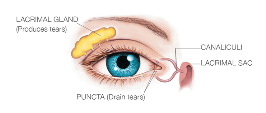
Like, the under production of tears. Or maybe the tear quality is just not good enough. And often, a combination of both. This is termed tear film instability. Causes for this instability are Meibomian Gland dysfunction, Corneal Surgery, Contact Lens wear, seasonal allergies, Systemic Diseases and long term use of certain eye drops (anti glaucoma).

Why are tears important?

A smooth layer of tears is essential for sharp vision. If the tear film has patchy areas where it has dried out, the light that hits the eye is scattered and does not focus clearly. A patchy, poor quality tear film also leads to discomfort and irritation.

What are the symptoms of Dry eye syndrome?

  1. Burning sensation and itching (Specially when exposed to increased evaporative conditions like heat, AC, prolong reading).
  2. Pricking sensation/ Foreign body sensation.
  3. Blurring of vision (especially at the end of the day).
  4. Redness and watering of eyes.
  5. Photophobia.
  6. Symptoms persists even after removing Contact lenses.

Patient’s experience of the disease may be far more uncomfortable than it appears to a clinician when examining the clinical sign.

Who is at risk of developing Dry eye syndrome?

  • Persons over 60 years of age
  • Women in general have higher chances
  • People who frequently use computer and video screens
  • Lack of sleep and excessive stress
  • Users of certain medications like anti-glaucoma drops, antihistamines and anti-depressants
  • Long term contact lens users

How is Dry eye syndrome treated?

First the cause of decreased tear film is identified. Depending on the cause and severity, Ophthalmologist will advise the course of treatment. Treatment options are:

Artificial tears

  • Artificial tears are eye drops used as a 
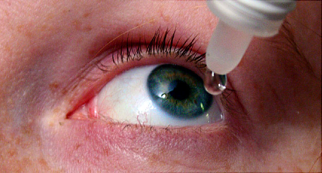
replacement for watery tears. Gels and ointments are also available, and are thicker than eye drops. (Gels usually last longer in the eye and need to be put in less frequently, but can blur the vision.)
  • Preservative-free eye drops are recommended for use with contact lenses, or if they are needed more than four times a day.
  • Expressing natural oily tears to the ocular surface by daily hot eyelid massage can reduce the frequency of artificial tear drops required

Antiglaucoma drops

  • Switching to preservative-free glaucoma eye drops is recommended for patients with glaucoma who also have dry eye syndrome.

Anti-inflammatory agents

  • Steroid eye drops or topical ciclosporin eye drops may be prescribed by an ophthalmologist if the dry eye condition has led to inflammation of the surface of the eye

Prevention of excessive drainage

  • When there is lack of watery (aqueous) tears, punctal plugs can be inserted into the tear duct (puncta) to stop tears from draining away and keep them on the surface of the eye for longer. It is a reversible procedure.
  • If the plugs help your dry eye but fall out, then punctal cautery can be considered. This is a permanent procedure.

How to prevent dry eye syndrome?

  1. Drink lots of water, as this helps improve tear production
  2. Get enough sleep each night
  3. Avoid alcohol or spicy foods, as these can cause meibomian oil gland blockage
  4. Avoid smoking or smoky areas as this makes the eyes drier
  5. Wear glasses or sunglasses on windy days to protect your eyes from the wind
  6. Avoid air-conditioned environments and draughts near your face, and consider using a humidifier to put more water into the air
  7. Remember to blink more frequently when using a computer screen or reading, and look far away (20 feet) from the screen for a break every 20 minutes, for 20 secs (20-20-20 rule)


Cataract and Its treatment– Everything you need to know!

Cataract & Its treatment– Everything you need to know!

What is a cataract?
The lens is a clear disc that receives light through the pupil and focuses it on the retina and
helps produce a clear image. Lens is mainly composed of water and proteins. As we age these
proteins may begin to clump together and cloud the lens. This clouding is called cataract.

What are the causes of Cataract?
Apart from age related, cataracts can also be caused by ultraviolet radiation, obesity, smoking,
eye injury, high myopia, steroid medications and family history.
What are the Symptoms of Cataract? How is cataract diagnosed?
Symptoms include blurred vision, double vision, increased glare, appearance of halos around lit
objects, and faded appearance of colors. It is diagnosed at a comprehensive eye exam.

Blurred Vision Glare at night

When should I have cataract surgery?
A cataract needs to be removed only when vision loss interferes with your daily activities, such
as driving, reading, or watching TV. You and your doctor can make this decision together. Once
you understand the benefits and risks of surgery, you can make an informed decision. In most
cases, delaying cataract surgery will not cause long-term damage to your eye. You do not have
to rush into surgery.

At times a cataract may need to be removed even if it does not cause visual problems. For
instance, a cataract extraction is necessary if it prevents examination or treatment of retinal
disorders (age-related macular degeneration or diabetic retinopathy) and Glaucoma (Lens
induced).

Why should I have cataract surgery?
Cataract-related vision loss is reversible with cataract surgery, which is one of the safest
surgeries in Ophthalmology.

What should I expect on the day of surgery?
Before surgery you will be given eye drops to dilate the pupil, the area around your eye will be
washed and cleansed and a local anesthetic maybe injected in the area surrounding your eye.
You may be offered a sedative to help you relax. Cataract surgeries generally last less than 30
minutes.

What does cataract surgery involve?
Common method used is Phacoemulsification. In this procedure the surgeon looks through an
operating microscope and makes three small incisions, less than one inch long, in or near the
cornea. An opening is made in the capsule of the lens and an ultrasound probe is used to break
the lens into tiny fragments. These fragments are aspirated out with the probe. A customized,
transparent artificial prescription lens called an intraocular lens made of silicone or acrylic
compounds, is inserted. The incisions are so small stitches are generally not required.

Is there any further advance in technique?
Femtosecond Laser Assisted Cataract surgery (FLACS) is a recent development in cataract
surgery. Femtosecond Laser is utilized to make the incisions, the opening in the lens capsule as
well as for fragmentation of the cataract nucleus. FLACS can offer greater level of precision and
to some extent can increase patient safety and outcome especially in cases like Fuch’s and
subluxated lens.

Cost of this procedure is significantly high.

What happens after the surgery?
Patient improves notably almost immediately after the surgery. After the procedure your eye
maybe patched for a day. Cataract surgery is an outpatient procedure and you can be
discharged on the same day. You can resume normal activities within a day or two. Exposure to
bright light may initially be uncomfortable, do not plan on driving immediately following the
procedure.

You may feel mild pain for a day or two which will alleviate with the eye drops and painkillers
prescribed.

Is there any alternative to surgery?
No. Don’t delay in getting an eye-exam.

Dr. Vidhi Majithia
Specialist Ophthalmology

TO know more http://bit.ly/30eGiE7
For Appointment : Call +971 4 406 3000 /02


Cataract Its treatment– Everything you need to know!


×