Orthopedic Care - Best Private Hospital in Dubai Al Mankhool | IMH Dubai

Sports Injury: When Should You See a Doctor

Sports injury hardly spares anyone. You may be a regular fitness freak or a weekend runner- sports injuries are bound to happen at some point or the other. You may be in two minds about seeing a doctor when such an injury happens. In this article, we will provide you few guidelines to help you decide when you should seek medical help in case of a sports injury.

Consistent Pain and Aches:

Regular pain and aches pose a constant discomfort in your daily life. Every time you go for that jog and run, that constant pain in the ankle is no double a headache. When the pain becomes a part a parcel of your regular exercise, you might consider seeking the help of a medical professional. Sometimes, the only time you will feel the pain is during and immediately after you exercise or engage in any outdoor games. Chances are, it will not impact your daily routine activities.

This type of injury is more commonly known as an overuse injury. So far you do not notice any swelling, bruise, limited physical activity or locking in your joint, you would be good without seeing a doctor. While caring for overuse injury, you have to be careful and watch out for anything serious. Put an ice pack, brace it while exercising, take painkillers like Ibuprofen and let it rest for a while when you can. In spite of all these methods, if your overuse injury does not heal when you would expect it to or if any of the above mentioned symptoms surface or persist, it is time to seek medical help.

Serious Injuries:

In the middle of a football match if you injure yourself or strain your ankle while running, it is not a regular overuse injury. If such an injury occurs, you might have swelling, discoloration, pain, numbness and you will be unable to walk normally. There might also be bleeding or a bone misalignment. In case of a serious injury like this, it is advisable to see a doctor right away. The doctor will do a physical examination and for proper diagnosis will order X-rays. Depending on the type of injury, the doctor might also provide a cast, splint or boot. Some serious injuries may also need Physio Therapy for proper healing.

Which Doctor to See:

Sometimes your injury will land you straight to the emergency room, but for other times you can choose the doctor you want to see for your sports injury. If you are a part of an organized sports program and play in a football, cricket or basketball team, you may have a team physician to attend to you. Otherwise you can choose the doctor whom you would like to see.

Although most family physicians are our first choice for basic injury care, it is advisable to consult an Orthopedic Surgeon who specializes in Sports Medicine.

When you are living an active life, injuries are bound to happen. It is important to determine which kind of injury needs medical treatment and whom to approach so that you are better prepared for an emergency.

At IMH, we have a highly qualified team of Orthopedic surgeons managing all kinds of orthopedic cases and sports medicine. The department has an outpatient clinic and 24/7 emergency services. Our Physiotherapy department has dedicated and highly qualified physiotherapists who work with the department. We would be happy to assist you in case if you need our support! For fresh injury and revision ligament  surgery you can meet our expert.

Dr. Gaurav Prakash Singh, Specialist Orthopedic Surgeon


Everything You Need to Know About Minimally Invasive Spine Surgery (MISS)

For as long as humans have lived, there has been the need to treat ailments — including spine issues. Thus, traditional methods of treating spinal issues have existed for thousands of years, even before the first-ever recorded operative spine surgery credited to Paul of Aegina in the 7th century. 

However, minimally invasive spine surgery (MISS) is still a relatively new method of treatment. It started becoming popular in the 1990s due to advancements in medical technology.

This piece will be looking at what minimally invasive spine surgery (MISS) is, its benefits, types, and how to prepare if you’re to undergo surgery. 

What is Minimally Invasive Spine Surgery (MISS)

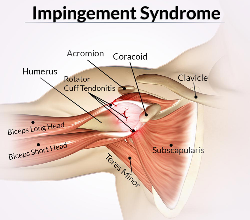
Minimally invasive spine surgery (MISS) is a surgical procedure performed to treat or correct spinal problems. Some of the most common spinal issues treated using MISS are scoliosis, herniated disc, spinal stenosis, degenerative disc disease, etc. 

The difference between MISS and the traditional open surgical method is that only small incisions are required to treat the patient with MISS. 

Thus, here are the five major advantages of MISS. 

Benefits of Minimally Invasive Spine Surgery (MISS)

Little Scars: Because Miss requires minimal incisions, patients get only small scarring and lesser risk of muscle damage. 

Fast recovery: Due to minimal scarring, MISS’s healing process is faster compared to traditional methods.

Reduced infection risk: The risk of infection is drastically reduced because MISS involves little muscle and tissue tearing. 

Minimal time at the hospital: Unlike regular spine surgeries, with minimally invasive spine surgery, you can go home within a few hours to 2 days after the surgery. 

Types of Minimally Invasive Spine Surgery (MISS)

Spinal fusion: Using minimally invasive spine surgery, spinal fusion; that is, the joining of two or more bones in the spine, can be done to stop movement between the bones. This is usually required to treat spinal ailments like degenerative disc disease, fracture, scoliosis, etc. The benefit of using MISS for this type of surgery is that it improves recovery and reduces the chances of infection.

Lumbar disc arthroplasty: This procedure is an alternative to the more popular spinal fusion surgery. It involves replacing a degenerated disk in the lower part of the spine. In this case, an artificial disk made of cobalt-chromium and plastic is used to replace the worn-out disk. 

Lumbar disc arthroplasty is a major surgery that involves dissecting muscles and tissues at the back. However, only a small incision is made between the lower abdominal muscles to get to the spine using the minimally invasive spine surgery method. Besides, MISS patients usually spend less time in the hospital post-operation. 

Corpectomy: This procedure is usually employed to relieve pressure caused by a spinal tumor, fracture, bone spur, stenosis, etc. It involves the surgical removal of a bone or parts of it in the neck, mid-back, or lower back.

Discectomy: This involves the surgical removal of materials pressing against the spinal cord or nerve root. Discectomy is usually employed in treating medical issues like bone spur, nerve root compression, sciatica, etc.

Foraminotomy: As the name suggests, this procedure is employed to treat issues related to the foramen. That is the tunnel in the back where nerve roots exit the spinal cord. Usually, foraminotomy is done to widen the foramen to relieve the pains and muscle weakness caused by nerve root compression. Other conditions treated with this procedure include herniated disc, bone spur, foraminal stenosis, etc. 

Laminectomy: Laminectomy involves the surgical removal of the lamina, the backbone protecting the spinal canal. This procedure is employed when there is a need to relieve spinal cord pains caused by spinal stenosis, bone spur, or herniated disc. By using minimally invasive spine surgery, this procedure can be completed within a few hours. The patients can also go home the same day. 

Kyphoplasty: This procedure is employed to restore a fractured vertebra to its original shape. This procedure is usually necessary when there is a compression fracture in the spine. Cancer, injuries, or osteoporosis; the thinning of the bones can cause such issues. 

Spinal instrumentation: Spinal instrumentation is employed in minimally invasive spine surgeries to prevent movement in specific segments of the spine. It’s usually done after the surgery to ensure proper fusion and quicker healing post-operation.

Spinal Disorders Treated Using the MISS Method

Herniated discs: This is one of the causes of back pains experienced by adults. Herniated discs occur when some of the inner core of an intervertebral disc breaks through its protective outer layer. This often causes back pain and inflammation of nerves surrounding the disc. 

Scoliosis: It’s mostly common among children, but adults can also develop the condition. Scoliosis is an abnormal sideward curvature that may lead to uneven shoulders, hips, or waist. Mild scoliosis may require regular monitoring but no surgery. However, if the condition worsens, an orthopedic doctor can employ MISS to prevent permanent deformity. 

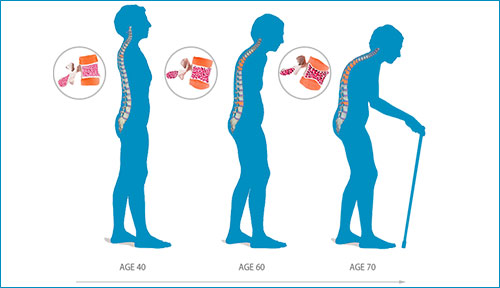
Degenerative disc disease: This disease occurs when the intervertebral discs stiffen and lose strength and shape. When this happens, the discs also lose the flexibility to absorb and distribute forces with spinal movements. Degenerative disc disease (DDD) is common in older adults. If left untreated, it could increase the chances of herniated discs.

Spinal stenosis: This occurs when the spaces within the spine are compressed or narrowed. This condition can cause numbness, muscle weakness, pain, tingling sensation at the back and neck, and sometimes in the arms and legs. In some cases, there are mild to no symptoms. In severe cases, spinal surgery may be required to create more openings for the spinal cord.

How To Know If You Need a Minimally Invasive Spine Surgery

It’s important to note that not every back pain requires MISS or any other kind of surgery.

If you are experiencing minor back pains, it may be because of your sitting posture, lack of sleep and exercise, etc. So firstly, try to correct such habits. 

That said, it’s essential to seek medical attention immediately if you are experiencing continuous pains or irritation in your neck and back areas. 

How to prepare for a MISS surgery?

Ideally, once your orthopedic doctor confirms that you need to undergo a MISS or any spine surgery, he or she will guide you on how to prepare yourself for it. 

Generally, you’ll be required to make some lifestyle adjustments like quitting smoking (if you’re a smoker), exercising regularly, eating healthy, and stopping certain medications. Such changes are necessary to minimize risks and quicken post-operation recovery.

If you’ve more questions about minimally invasive spine surgery or any other spine-related disorders and treatments, feel free to reach out to us. Also, you can book an appointment to see one of our orthopedic doctors in Dubai. 

Osteoporosis: Causes, Symptoms & Prevention


What Is Osteoporosis?

Osteoporosis is a common disease that makes bones thinner, which makes them more likely to break. These fractures can lead to different health problems, like pain, bent posture, or trouble moving around. Many people lose bone gradually over many years. There are no symptoms that tell you that it’s happening. But it is possible to treat osteoporosis with medications and healthy lifestyle choices. If you make those good habits part of your life early on, you can prevent bone loss and lower the chances you’ll break a bone. It is easily reversible.

What Causes Osteoporosis?

Human body constantly breaks down old bone and rebuilds it. This process is called remodeling. As you grow up, your body builds more bone than it removes. During childhood, your bones become larger and stronger. Peak bone mass happens when you have the most bone you will ever have, usually in your early to mid-30s. At a certain age, the bone remodeling process changes. New bone comes in at a slower rate. This slowdown leads to a drop in the amount of bone you have. When bone loss becomes more severe, you have osteoporosis.

Osteoporosis

Osteoporosis Symptoms

Osteoporosis usually doesn’t cause any symptoms. But after many years, you may notice signs like back pain, a loss of height, or a stooped posture. For some people, the first sign they have the disease is a broken bone, usually in the spine or hip unless you get early screening done, or it gets noticed when you get an X ray for some other reason , the initial sign of osteoporosis is fractured or broken bone .
If osteoporosis becomes severe, the normal stress on bones from sitting, standing, coughing, or even hugging can cause painful fractures. After the first fracture, you’re more likely to get more.
For some people, the pain from a fracture may get better as the bone heals. But others will have long-lasting pain. You may feel stiff and have trouble being active.

Things that raise your chances of getting osteoporosis include:

  • Family history: Osteoporosis seems to run in families. If your mother had a hip or spine fracture due to osteoporosis, chances are you’re more likely to get the disease.
  • Gender: Women are four times more likely than men to get osteoporosis.
  • Age: Though anyone can have osteoporosis, your chances go up with age. Women over 50 are most likely to get it. The older you are, the more likely you are to have fractures.
  • Bone structure and body weight: Petite and thin women have higher chances for the disease, too. Weight loss after age 50 in women also seems to raise the chance of hip fractures, while weight gain lowers it. Small-boned, thin men have a greater chance of getting osteoporosis than men with larger frames and more body weight.
  • History of fractures: Having one fracture due to osteoporosis means you’re likely to get more.
  • Smoking: Studies show that cigarette smokers (past or current) have lower bone mass and higher fracture risks. Women who smoke have lower levels of the hormone estrogen — a key part of bone health.
  • Medications: Some medications may make you more likely to have the disease. These include long-term use of steroids (prednisone), thyroid drugs, anti-seizure medicine, antacids, and other drugs.


How can I prevent osteoporosis before it starts?

Osteoporosis is a largely preventable disease. Prevention should start early. Getting enough calcium and vitamin D as a child and teenager can dramatically cut your risks of developing osteoporosis later in life. Even if you’re an adult, eating a healthy diet, getting enough calcium and vitamin D, exercising, and avoiding unhealthy lifestyle habits such as smoking and excessive drinking, may help prevent osteoporosis. Walking at least three times per week has proved to help reduce the progression of osteoporosis. After menopause, women who have osteopenia, or thinning of the bones, and who have a high probability for a future fracture from osteoporosis can consider drug therapies to prevent bone loss and reduce their risk of osteoporosis. To learn about osteoporosis prevention techniques, talk to your health care provider.
Am I getting enough calcium — and how much is too much?

The amount of calcium you need depends on your age. The Institute of Medicine recommends the following:

  • Adolescents should get 1,300 milligrams of calcium a day.
  • Adults from 19 to 50 years of age should get 1,000 milligrams of calcium a day.
  • Adult women over the age of 50 should get 1,200 milligrams of calcium a day
  • Adult men should receive 1,000 milligrams up to age 70 and 1,200 milligrams after age 70.
  • Read food labels and select foods that contain 10% or more of the Daily Value for calcium. When food shopping, look for terms such as “high in calcium,” “fortified with calcium,” “calcium-rich,” or “excellent source of calcium.”
  • If you think you’re coming up short, talk to your health care provider about ways you can boost your calcium levels in your diet. Talk to your doctor about what is best for you.

What should I use after injury ? Cold or Heat?

Orthopedic Care DubaiInjury to the extremities and   trunk is quite common  which  almost everybody gets by normal activities or  by an accident like playing football, lifting heavy weight etc.. There seems to be the confusion about application of ice or hot pack  and how long one should use after the injury.

It is advised to  use ice in the acute stage of an injury (within the first 24-48 hours), or whenever swelling is develops. Ice helps to reduce inflammation and swelling by decreasing

Orthopedic Care in Dubai

blood flow to the area that is injured. One should  apply ice indirectly (not directly on the skin) for 10 to 20 minutes, remove the ice for at least 20 minutes, and repeat as necessary. Heat increases blood flow, which helps promote pain relief after inflammation and swelling subside. Heat is also used to assist in warming muscles up prior to exercise, any physical activity, or physical therapy.

Morning Stiffness & Back Pain

Waking up with back pain is rarely a serious problem. Even many people who are more or less pain free during the day still experience pain in the nights and while getting up.
morning stiffness & back pain
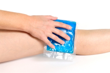
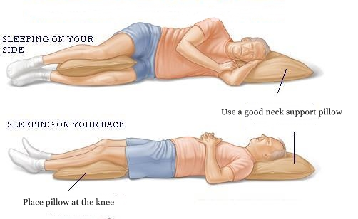
Recent research reveals that nearly one in three people experience back pain after a night’s sleep, No one really understands the reason for morning stiffness. Presumably, while the body is inactive, fluid leaks out from the small blood vessels and capillaries and the tissues become “waterlogged.” Then, if you try to move the part, the swollen tissues feel stiff until the motion pumps the fluid out through the lymph channels and the veins while most feel stiff or ache.

The average episode of morning stiffness lasts only about 10 to 15 minutes. The stiffness goes away as you move and warm up the joints and muscles. However, stiffness from rheumatoid arthritis may last more than an hour.
People often say, “I’m just getting old,” but old age alone does not cause morning joint stiffness. It is usually an indication of worn joints, muscle tightness, or inflammation from arthritis.When your lower back is stiff in the morning, muscles such as psoas, gluteus and hamstrings become tight overnight. This generates pressure on the joints resulting in pain. Weak abdominals also contribute to this pain as they do not allow adequate support for the lumbar spine

As your joints get older, the spongy cushion of cartilage begins to dry out and stiffen. The joint lining also produces less synovial fluid, which lubricates the joint. Weak muscles and stiff tendons also tend to tighten during sleep. Osteoarthritis, (the “wear and tear” kind), and rheumatoid arthritis, (which involves swelling and inflammation), both can trigger morning stiffness.

In some of the case, spinal morning stiffness is associated with lumbar disc disorder . The associations increases when we combined spinal morning stiffness with low back pain.

There are various causes and contributing factors for morning stiffness, and some of them are –
• Overweight and poor diet
• Too much exposure in a cold environment
• Poor posture while sleeping .

No question that the sleep surface can influence force into the soft tissues. If you don’t believe , try sleeping one night on the kitchen floor and you’ll realize it.

Remedies of morning stiffness

• Regular exercise
• Avoid stomach sleeping.
•Sleep position- Place a plush, supportive pillow underneath your knees, if you sleep on your back. This action flattens your back, removing a large arch from your lower back region. It can relieve pain in just a few minutes. If you are a back and side sleeper, you can use a supportive pillow and pull it under your knees or between your legs as you switch positions.Remedies of morning stiffness

•You can also place a small, rolled-up towel under the small of your back for extra support. You need a firm mattress (not too soft / not too hard). Your bed should form to the shape of your body

• Stretching and sitting up while in bed.- Think of stretching in the morning as you warm up for the entire day. These stretches are meant to lightly ease the body into movement, and upon completion, leave you feeling more awake and ready to get on with your day.

• Stretching in the morning will ease stiffness, soreness, and can help diminish chronic aches and pains in the body. Take time for yourself in the morning with yoga poses if you know, so you can be sure to enjoy the rest of the day!

•Drink enough water -. Make sure you’re well hydrated. This might seem obvious as soft tissues need water to remain as pliable as possible.

• Healthy diet
• Hot shower
•Learn techniques for stress management – Learning stress management techniques can assist you to sleep comfortably

•Supplements : You cannot reverse the effects of joint aging, and while certain medications can help manage arthritis pain and inflammation, stiffness can still occur.. Maintaining a healthy weight also can help. Finally, don’t be swayed by joint health supplements, such as glucosamine or chondroitin.. This is controversial because there have been studies that suggest Glucosamine and Chondroitin Sulfate don’t work for arthritic joints even though is chondroitin Sulfate is a disease modifying agent for osteoarthritis. It slows the progression of the disease and deterioration of the joint cartilage.

• Avoid painkillers, they don’t help morning stiffness.

Limb Lengthening

How Does It Work?

The limb lengthening process works by gradually growing new bone and soft tissues (skin, muscle, nerves, blood vessels, etc). This new growth is called tissue regeneration.

Bone and soft tissue regenerate when they are distracted (pulled apart) at a very slow rate of approximately 1 mm per day. If the rate of distraction is faster than this, bone may fail to form between the two ends of the bone that are being pulled apart. Then, soft tissues, such as muscle, may experience contracture (get too tight) or nerves may become paralyzed. If the rate of distraction is too slow, premature consolidation may occur (the bone may consolidate too soon), preventing the lengthening device from further pulling it apart.

There are many different lengthening devices used. The most common are external fixators, which are devices that attach to the bone by means of thin wires or thicker pins that have a screw threading at their attachment to the bone. There are also lengthening devices that are fully implanted inside the bone. These devices do not require external pins. The different devices are described separately.

There are two phases of lengthening until the bone is fully healed: the distraction phase and the consolidation phase. The distraction phase is the lengthening phase. After the desired length is obtained, the newly regenerated bone is still very weak because of lack of calcium within it. The hardening and calcification of this new bone is called the consolidation phase.

METHODS OF LENGTHENING:-

A variety of orthopedic devices are used to distract the bone and soft tissues. The decision regarding which device to use is an individualized one dependent upon the desired correction.

There are two general types of devices: external fixators and internal fixators. The external devices attach to the bone from outside the body by means of wires and threaded pins. The internal devices are implanted inside the body and lie on the bone or inside the marrow cavity of the bone.

The best known and most versatile techniques are with monolateral (one-sided or straight bar) external fixators (e.g., Orthofix, EBI) and circular external fixators (e.g., Ilizarov, TSF). External fixators are applicable to almost all cases. To shorten the time in the external fixator, combinations of the external fixator method with an internal nail to perform the lengthening over nail (LON) technique. However, this method is not applicable to all cases.

A fully implantable self-lengthening nail (ISKD) is also used. It has an internal mechanism that performs the lengthening and thereby eliminates the need for an external fixator. This method is available for certain cases.

External Fixation Alone
When only an external fixator is used, the fixator needs to remain in place for both the distraction and consolidation phases. If the fixator were removed at the end of the distraction phase, the new bone would collapse and re shortens. Therefore the external fixator needs to remain in place until the regenerated bone appears solid enough on the radiographs (x-rays). At that point, the device can be removed. Often, a cast is applied to temporarily protect the bone from breakage for an additional short time.

The total time in the external fixator can be estimated to be approximately 1 month for each centimeter lengthened in children and 1.5 to 2 months or longer per centimeter in adults.

Lengthening over Nail
Lengthening over Nail or LON was developed in order to decrease the external fixator treatment time, eliminate the need for post-removal casting, and decrease the risk of breakage of the new bone.

With LON, a metal rod is inserted into the bone along with the external fixator. The rod fits in the marrow cavity of the center of the bone, and the external fixator is applied around the peripheral part of the bone so that the external fixator pins do not come in contact with the metal rod. The bone is lengthened as described above.

After lengthening is completed, the patient goes back to the operating room for the insertion of special screws that lock the rod to the bone. With screws at both ends of the rod on opposite sides of the lengthening zone, the external fixator is no longer needed, and is removed during the same operation.

Therefore, the total external fixation treatment time is much shorter, and is equal to the distraction phase. This usually reduces external fixator time to less than half.

Fully Implantable Lengthening Nails and Prostheses
The most recent development is that of a fully implantable devices that can lengthen the limb from within. There is no need for an external fixator. This has many advantages, including no risk of pin infection, no muscle tethering by the pins, less pain, and better comfort. Unfortunately, , this method is mostly limited to older children and adults. Therefore, many patients cannot be treated by this method.

Leg length discrepancy correction

limb1

Bow leg correction

limb2

Impingement Syndrome

shoulder-impingement-syndrome-2

Shoulder impingement syndrome is also called as subacromial impingement, painful arc syndrome, supraspinatus syndrome, swimmer’s shoulder, and thrower’s shoulder.
It is associated with pain and some weakness when you raise your arm, and occurs when the tendons of the rotator cuff become inflamed when they pass through a narrow subacromial space.

In this syndrome, the rotator cuff tendon becomes trapped in the narrow subacromial space and repeatedly scrapes against the bone above(acromion) causing pain which becomes worse when you raise your arm over your head.

It may also be associated with persistent ache in your shoulder and pain at night.

Onset

This can start suddenly after an injury, or during middle age onwards it can come on gradually without any obvious cause.

Causes
1. Bony spurs under the acromion at the top of the subacromial space – this can narrow the space
2. swelling or thickening of the rotator cuff tendon – due to injury or inflammation
3.Bursitis- inflammation of the fluid-filled sac (bursa) that lies between the rotator cuff tendon and acromion
4. Calcific tendinitis – Calcification deposits within the rotator cuff tendon

Management Conservative

  • Rest
    Pain Killers – to ease the pain
  • Physiotherapy

After examination we will decide on referring you to a Physiotherapist to provide sessions.

Injections

A combination of Local anaesthetic and corticosteroid is injected which helps to reduce the inflammation. Further physiotherapy is recommended. Symptoms may recur.

impingement-syndrome

Surgical

Surgery
After considering these above modalities and if they aren’t effective, we will discuss with you the option of surgical management which involves an operation to widen the subacromial space in your shoulder, so the rotator cuff tendon is no longer rubbing against the bone above. This is known as subacromial decompression.

It can usually be done using a technique called arthroscopy, which is a type of keyhole surgery carried out under general anaesthetic, where special surgical instruments and a thin, flexible tube containing a camera are inserted through small cuts in your skin.

The recovery is faster and with less scarring than if a larger cut was made as is routinely done in Open surgery. Most people are able to go home early .

This technique can also be used to

  1.  remove any calcium deposits that have formed in the tendon in your shoulder
  2.  Repair the rotator cuff tendons if injured and many more.

Dr. Vijayakumar Kunthe,
MBBS, D.Ortho, DNB (Ortho), MNAMS, MRCS (Ed) (UK), Fellow Shoulder Surgery (UK)
Specialist Orthopedic Surgeon

Meniscal cartilage injuries

Meniscal cartilage injuries

The knee is commonly injured in sports, especially rugby, football and skiing. You may tear a meniscus by a forceful knee movement whilst you are weight bearing on the same leg. The classical injury is for a footballer to twist (rotate) the knee whilst the foot is still on the ground – for example, whilst dribbling round a defender. Another example is a tennis player who twists to hit a ball hard, but with the foot remaining in the same position. The meniscus may tear fully or partially. How serious the injury is depends on how much is torn and the exact site of the tear.

Meniscal tears may also occur without a sudden severe injury. In some cases a tear develops due to repeated small injuries to the cartilage or to wear and tear (degeneration) of the meniscal cartilage in older people. In severe injuries, other parts of the knee may also be damaged in addition to a meniscal tear. For example, you may also sprain or tear a ligament.

Meniscal cartilage does not heal very well once it is torn. This is mainly because it does not have a good blood supply. The outer edge of each meniscus has some blood vessels, but the area in the centre has no direct blood supply. This means that although some small outer tears may heal in time, larger tears, or a tear in the middle, tend not to heal.

What are the symptoms of a meniscal tear?
The symptoms of a meniscal injury depend on the type and position of the meniscal tear. Many people have meniscal tears without any knee symptoms, especially if they are due to wear and tear (degeneration).
• Pain. The pain is often worse when you straighten the leg. If the pain is mild, you may be able to continue to walk. You may have severe pain if a torn fragment of meniscus catches between the tibia and femur. Sometimes, an injury that you had in the past causes pain months or years later, particularly if you injure the knee again.
• Swelling. The knee often swells within a day or two of the injury. Many people notice that their knee is slightly swollen for several months if the tear is due to degeneration.
• Knee function. You may be unable to straighten the knee fully. In severe cases you may not be able to walk without a lot of pain. The knee may lock from time to time if the torn fragment interferes with normal knee movement. Some people notice a clicking or catching feeling when they walk. (A locked knee means that it gets stuck when you bend it and you can’t straighten it without moving the leg with your hands.)
Note: a ‘clicking’ joint (especially without pain) does not usually mean you have a meniscal tear.
For some people, the symptoms of meniscal injury go away on their own after a few weeks. However, for most people the symptoms persist long-term, or flare up from time to time, until the tear is treated.

How is a meniscal tear diagnosed?

• The story and symptoms often suggest a meniscal tear. A doctor will examine the knee. Certain features of the examination may point towards a meniscal tear.
• Your doctor may sometimes advise an X-ray of the knee – but this is often not necessary. An X-ray will not show cartilage tissue, but it can check for any bone damage which might have also occurred with the injury.
• The diagnosis can be confirmed by an MRI scan of the knee
What is the treatment for a meniscal tear?
When you first injure your knee the initial treatment should follow the simple PRICE method:
• Protect from further injury.
• Rest (crutches for the initial 24-48 hours).
• Ice (apply ice (wrapped in a towel, for example) to the injured area for 20 minutes of each waking hour during the first 48 hours after the injury).
• Compression (with a bandage, and use a knee brace or splint if necessary).
• Elevation (above the level of the heart).
These actions, combined with painkillers, help to settle the initial pain and swelling.

Surgery
If the tear causes persistent troublesome symptoms then an operation may be advised – although evidence for the benefit of some types of surgery is variable. Most operations are done by arthroscopy (see below). The types of operations which may be considered include the following:
• The torn meniscus may be able to be repaired and stitched back into place. However, in many cases this is not possible.
• In some cases where repair is not possible, a small portion of the meniscus may be trimmed or cut out to even up the surface.
• Sometimes, the entire meniscus is removed.
• Meniscal transplants have recently been introduced. The missing meniscal cartilage is replaced with donor tissue, which is screened and sterilised much in the same way as for other donor tissues such as for kidney transplants. These are more commonly performed in America than in the UK.
• There is a new operation in which collagen meniscal implants are inserted. The implants are made from a natural substance and allow your cells to grow into it so that the missing meniscal tissue regrows. This is not yet available at all hospitals.

Arthroscopy

This is a procedure to look inside a joint by using an arthroscope. An arthroscope is like a thin telescope with a light source. It is used to light up and magnify the structures inside a joint. Two or three small (less than 1 cm) cuts are made at the front of the knee. The knee joint is filled up with fluid and the arthroscope is introduced into the knee. Probes and specially designed tiny tools and instruments can then be introduced into the knee through the other small cuts. These instruments are used to cut, trim, take samples (biopsies), grab, etc, inside the joint. Arthroscopy can be used to diagnose and also to treat meniscal tears. Following surgery, you will have physiotherapy to keep the knee joint active (which encourages healing) and to strengthen up the surrounding muscles to give support and strength to the knee.


Dr. Mohamed Kandil

M.B,B.CH, Msc (Ortho), Dip.Sports Medicine
(UK),Fellowship (Germany)

Specialist Orthopedic Surgeon

Trigger Finger – Things to Know

Trigger finger is a painful condition that causes the fingers or thumb to catch or lock when bent. In the thumb its called trigger thumb.

The problem

    • Painful condition characterised by:

    • Inability to bend fingers freely
    • Caused by inflammation and thickening of the annular pulleys of the flexor tendons
    • Disorder characterised by inability to extend the flexed fingers
    • Painful and very common

download

Features

Women above 45 are commonly affected Common in patients with rhuematoid athritis The ring and middle fingers are most commonly affected

Treatment

  • Steroid injection

images

Often neglected cause of numbness of hands -Carpal tunnel syndrome

Carpal tunnel syndrome is a very common,easily treatable condition affecting middle aged people commonly.The condition is characterized by feeling of tingling and numbness in the hand.Sometimes there will be pain instead of the typical numbness.
download-11

THE PROBLEM

Compression of MEDIAN NERVE in the carpal tunnel
theproblm

carpal tunnel syndrome more commonly affects FEMALES in the age group 30 to 60 years .
men involved in HEAVY DRILLING ACTIVITIES and using VIBRATING MACHINERY are also susceptible.
obesity,smoking,alchoholism,rhuematoid arthritis and other inflammatory arthritis,hypothyroidism,diabetes mellitus
amyloidosis rarely affects children

CLINICAL SYMPTOMS
cli

NUMBNESS in the outer three and a half fingers
num

weakness of the thumb
decreased grip strength
wasting of thenar muscles
phalens test positive
durcums test positive
tinels sign

INVESTIGATIONS
Nerve conduction study
MRI
CT

TREATMENT
STEROID INJECTION AND NIGHT SPLINT – very early cases

CARPAL TUNNEL RELEASE SURGERY – later stages

strer


×